Eye Care Technology at International Eye Hospital, Lahore

Diagnosis is only as good as the equipment behind it. IEH Gulberg runs the most advanced ophthalmic platforms available in Pakistan — German, Swiss and Japanese systems used by leading centres worldwide, installed and calibrated here in Lahore.

  • Germany
  • Switzerland
  • Japan
  • USA
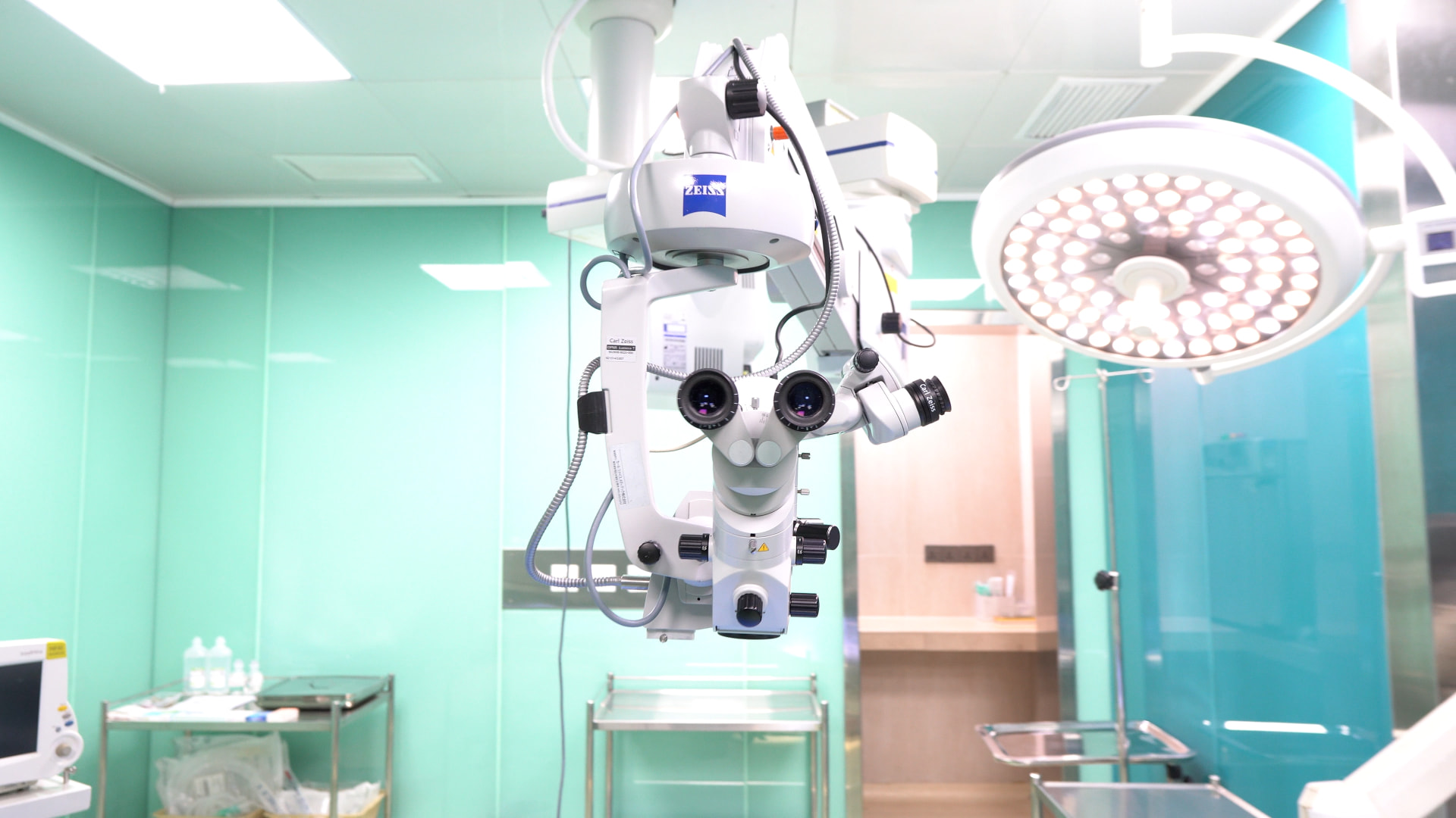
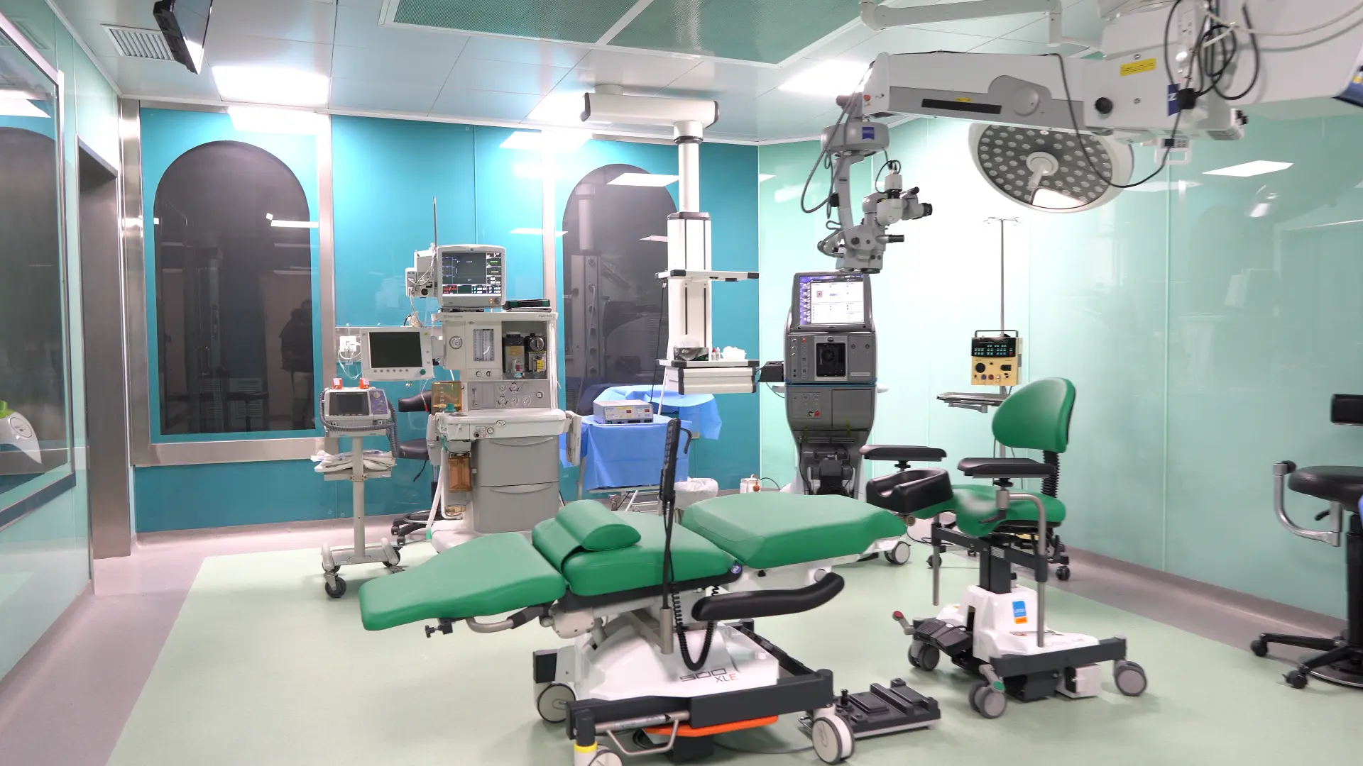
ZEISS OPMI Lumera T surgical microscope in the executive operation theatre at International Eye Hospital, Gulberg Lahore

Signature Platform

ZEISS OPMI Lumera T Surgical Microscope

The Lumera T is the microscope our cataract and retinal surgeons operate under every day, and its Stereo Coaxial Illumination produces a true-to-life red reflex that makes the lens capsule visible as it really is rather than as a guess. That stereoscopic clarity and depth of field let the surgeon judge tissue planes millimetre by millimetre through the most delicate steps. Because the illumination holds steady from first incision to lens implantation, nothing about the view changes at the moment precision matters most.

  • Stereo Coaxial Illumination for a brilliant, even red reflex
  • Superb detail resolution of capsule and vitreous structures
  • Integrated video documentation of every procedure
  • Floor-stand stability with fine motorised focus control
See the technology in detail
“The most advanced ophthalmic equipment in Pakistan — installed at IEH Gulberg.”
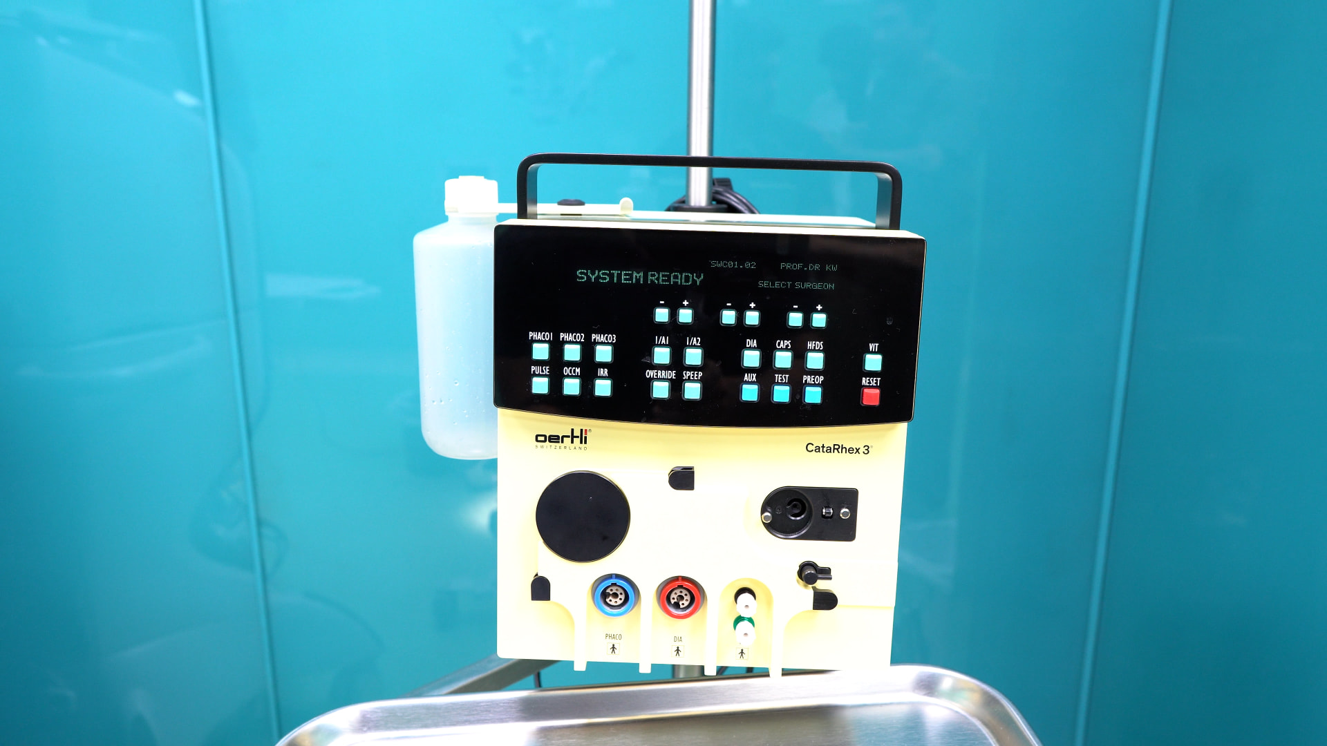
Oertli CATARHEX Swiss phacoemulsification console used for cataract surgery at International Eye Hospital, Gulberg LahoreSwitzerland

Oertli CATARHEX Cataract Surgery System

A leading Swiss phacoemulsification platform for cataract removal, built around fluid control and gentle energy delivery to protect delicate eye tissue during surgery.

  • Precise phacoemulsification control
  • Reduced energy delivery to the eye
  • Stable anterior chamber during surgery
  • Faster, safer cataract removal
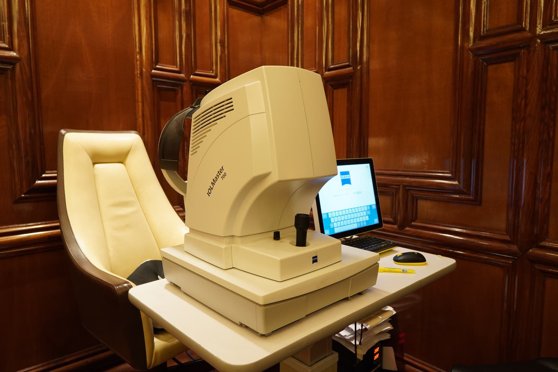
ZEISS IOL Master 700 swept-source biometer measuring the eye before lens implantation at International Eye Hospital LahoreGermany

ZEISS IOL Master 700 Optical Biometry

Swept-source optical biometry for cataract and lens surgery planning — measuring the eye with high precision to select the correct intraocular lens power before surgery.

  • Non-contact, high-precision measurement
  • Full eye length scan in seconds
  • Improves intraocular lens accuracy
  • Reduces post-surgery refractive surprises
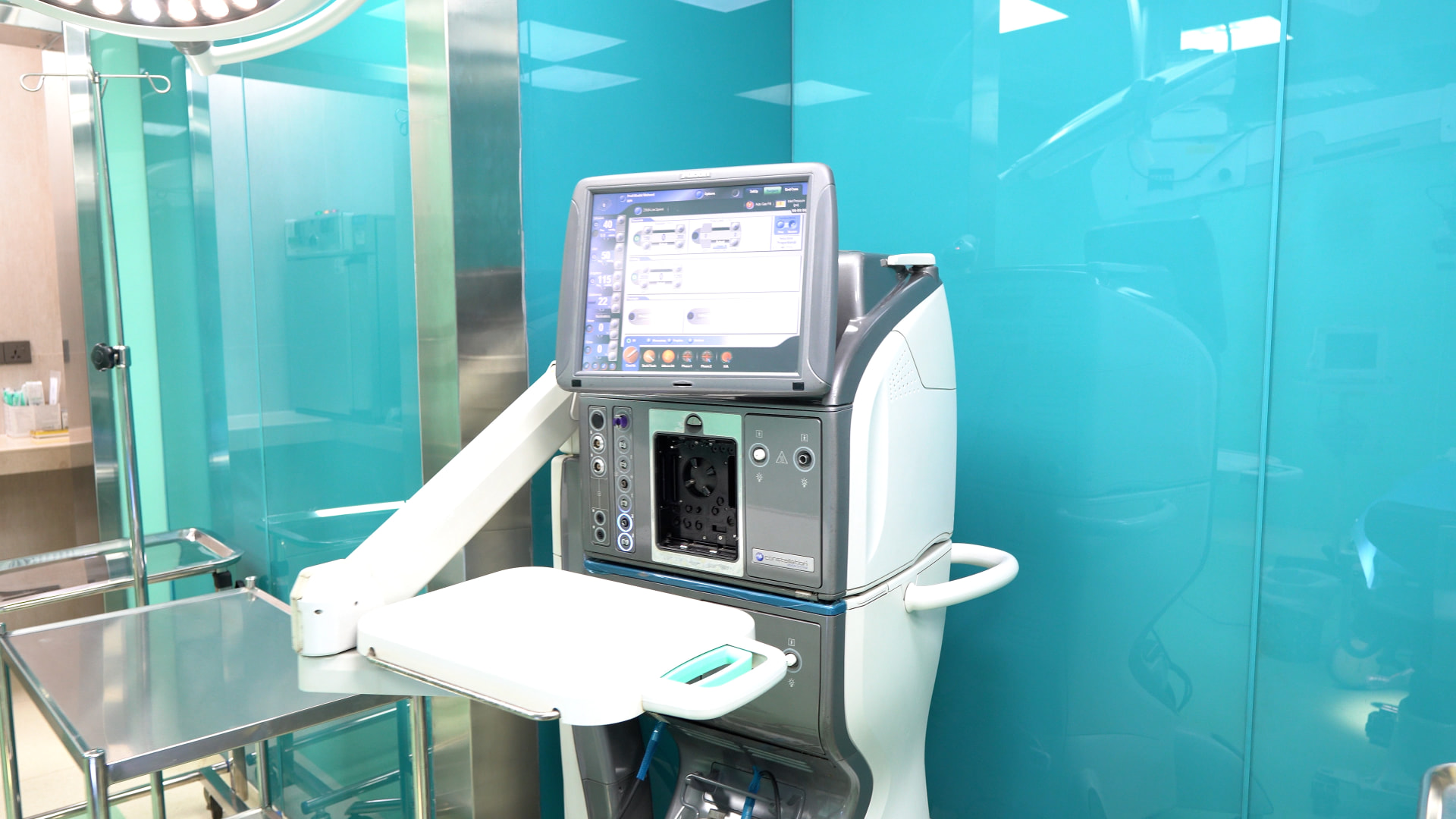
Constellation Vision System vitrectomy platform used for retinal detachment surgery at International Eye Hospital LahoreUSA

Constellation Vision System for Vitreoretinal Surgery

An advanced vitrectomy platform for complex retinal surgery, giving surgeons precise control over fluid dynamics and cutting speed during procedures like retinal detachment repair.

  • High-speed, precise vitreous cutting
  • Stable intraocular fluidics
  • Suited to complex retinal procedures
  • Reduces surgical trauma to the eye
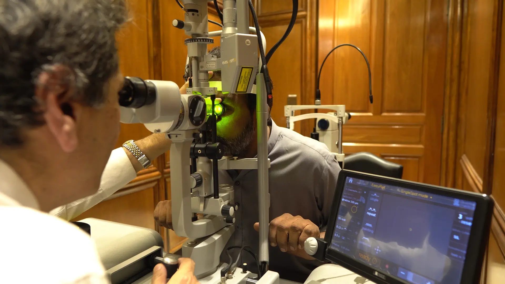
Subliminal yellow wavelength retinal laser available exclusively at International Eye Hospital, Gulberg LahoreExclusive to IEH in LahoreAvailable exclusively at IEH

Subliminal Yellow Laser Retina Treatment

A retinal laser platform using sub-threshold yellow wavelength technology, delivering treatment with less thermal spread to surrounding tissue than conventional argon lasers.

  • Sub-threshold, tissue-sparing delivery
  • Reduced thermal damage to the retina
  • Yellow wavelength targets vessels precisely
  • Available in Lahore only at International Eye Hospital
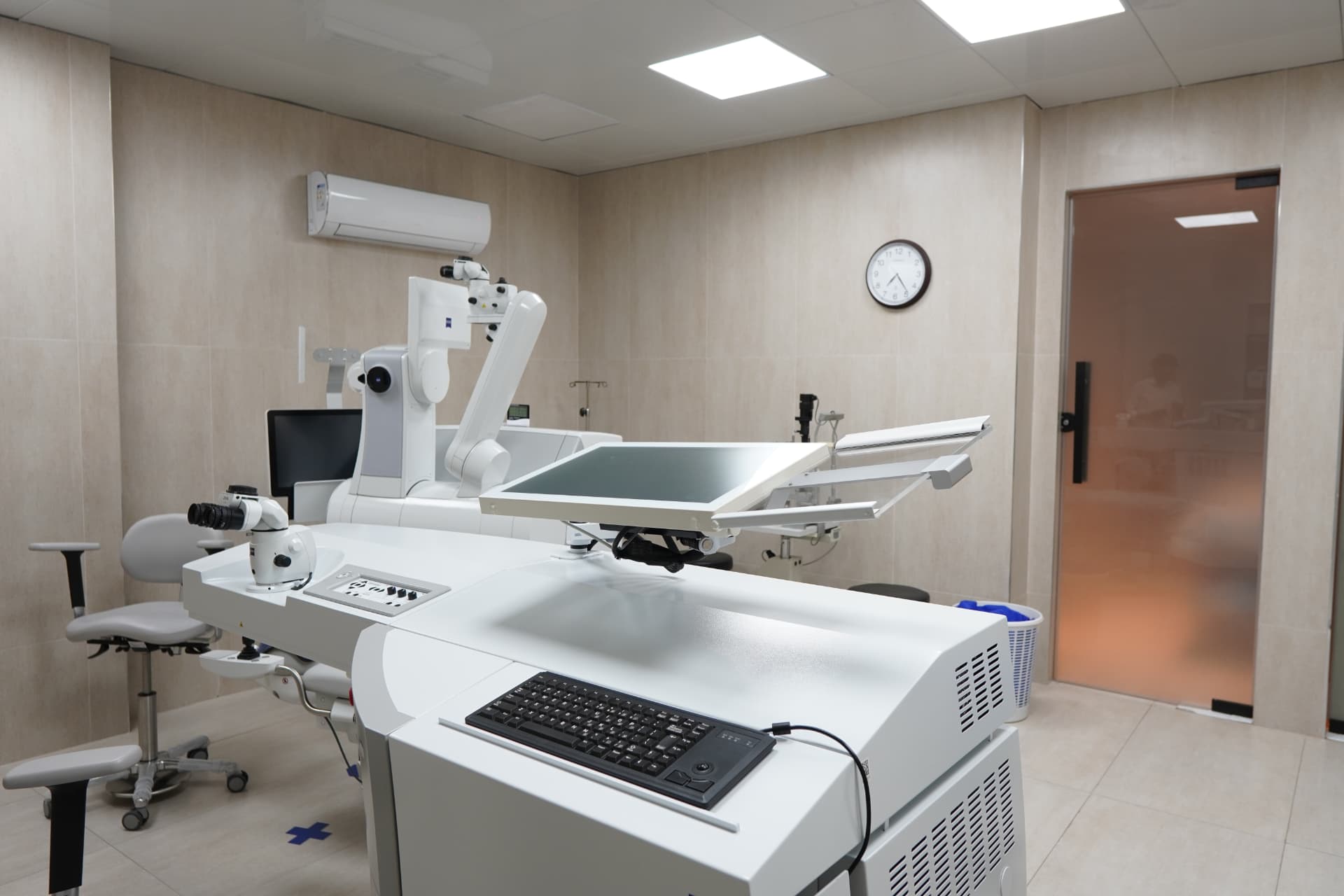
ZEISS VISUMAX 800 femtosecond laser used for SMILE Pro vision correction at International Eye Hospital, Gulberg LahoreGermany

ZEISS VISUMAX 800 — SMILE Pro Femtosecond Laser

The VISUMAX 800 is the fastest femtosecond platform ZEISS has built, running at 2 MHz to complete a SMILE Pro lenticule in under 10 seconds per eye. It is the engine behind flapless laser vision correction at our Gulberg hospital.

  • 2 MHz pulse frequency, under 10 seconds of laser per eye
  • CentraLign centration on the patient's visual axis
  • OcuLign cyclotorsion alignment for accurate astigmatism correction
  • Flapless keyhole technique preserving corneal biomechanics
ZEISS OPMI Lumera T surgical microscope in the executive operation theatre at International Eye Hospital, Gulberg LahoreGermany

ZEISS OPMI Lumera T Surgical Microscope

The OPMI Lumera T gives our cataract and retinal surgeons stereo red-reflex illumination with exceptional depth of field. Better visualisation of the capsule and vitreous translates directly into safer surgery.

  • Stereo Coaxial Illumination for a brilliant, even red reflex
  • Superb detail resolution of capsule and vitreous structures
  • Integrated video documentation of every procedure
  • Floor-stand stability with fine motorised focus control
Corneal cross-linking machine delivering calibrated UV-A treatment for keratoconus at International Eye Hospital LahoreSwitzerland

Corneal Cross-Linking (CXL) System

Our cross-linking system delivers a calibrated UV-A dose to riboflavin-soaked corneal tissue, stiffening collagen to arrest keratoconus. Dose accuracy is what separates effective treatment from under-treatment.

  • Calibrated UV-A emission with precise dose control
  • Supports standard, accelerated and epi-on protocols
  • Uniform beam profile across the treatment zone
  • Adjustable spot size for targeted corneal treatment

What we use it for

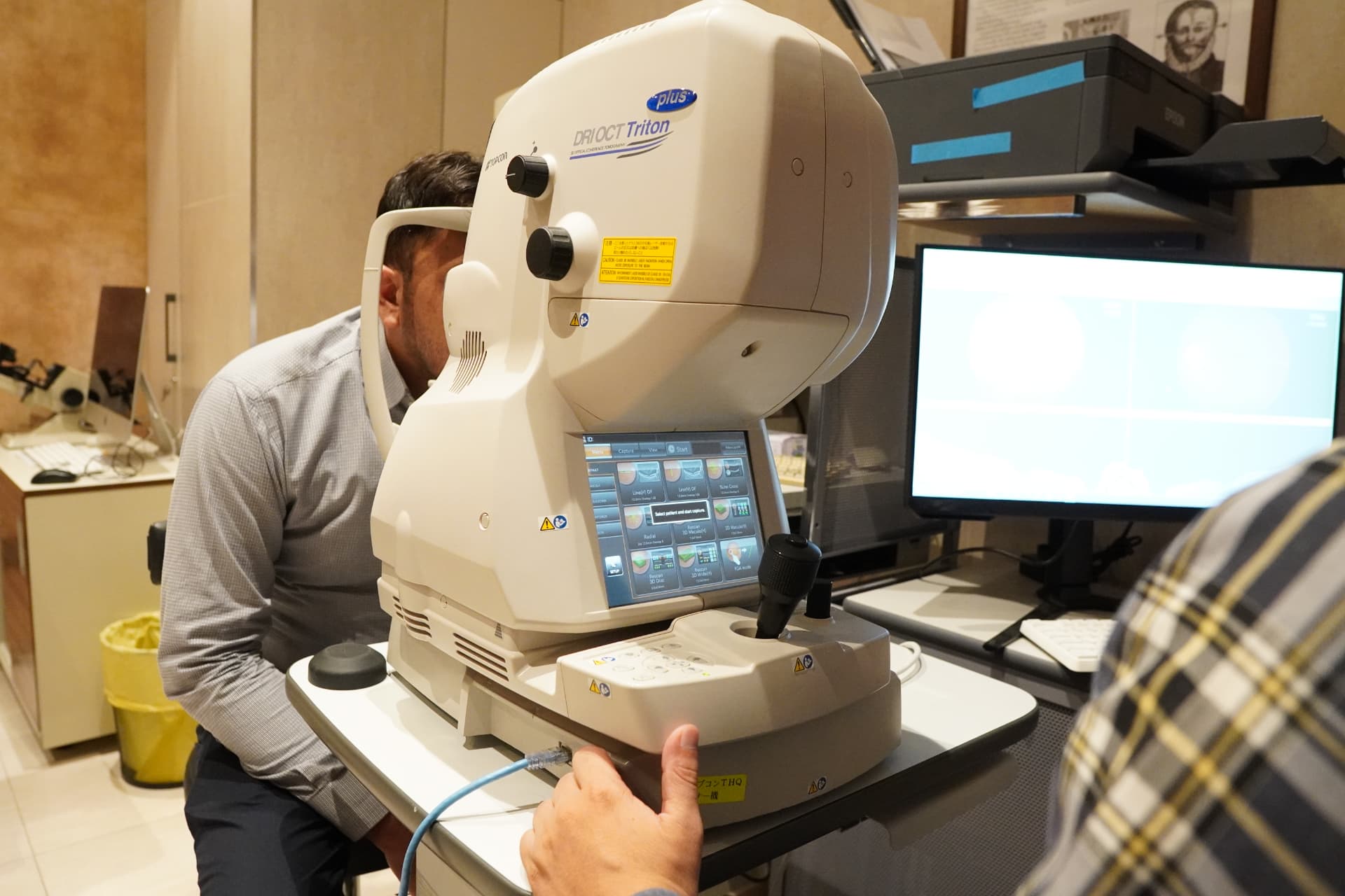
OCT scanner producing micron-resolution retinal and optic nerve images at International Eye Hospital LahoreGermany

Optical Coherence Tomography (OCT) Scanning

OCT produces cross-sectional scans of the retina and optic nerve at micron resolution, revealing fluid, membranes and nerve thinning invisible on examination alone. It underpins our diabetic, macular and glaucoma services.

  • Micron-resolution cross-sections of retina and macula
  • Retinal nerve fibre layer analysis for glaucoma tracking
  • Quantifies diabetic macular oedema before and after treatment
  • Progression analysis comparing scans over time
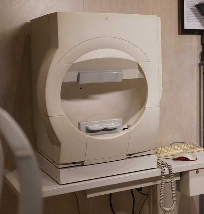
Automated visual field perimeter used for glaucoma monitoring at International Eye Hospital LahoreGermany

Automated Visual Field Perimetry

Automated perimetry maps your field of vision point by point to detect the peripheral loss caused by glaucoma and neurological disease. Repeat testing shows whether treatment is holding the disease steady.

  • Threshold testing across central and peripheral field
  • Reliable detection of early glaucomatous defects
  • Statistical progression analysis across visits
  • Screening protocols for driving and occupational needs

What we use it for

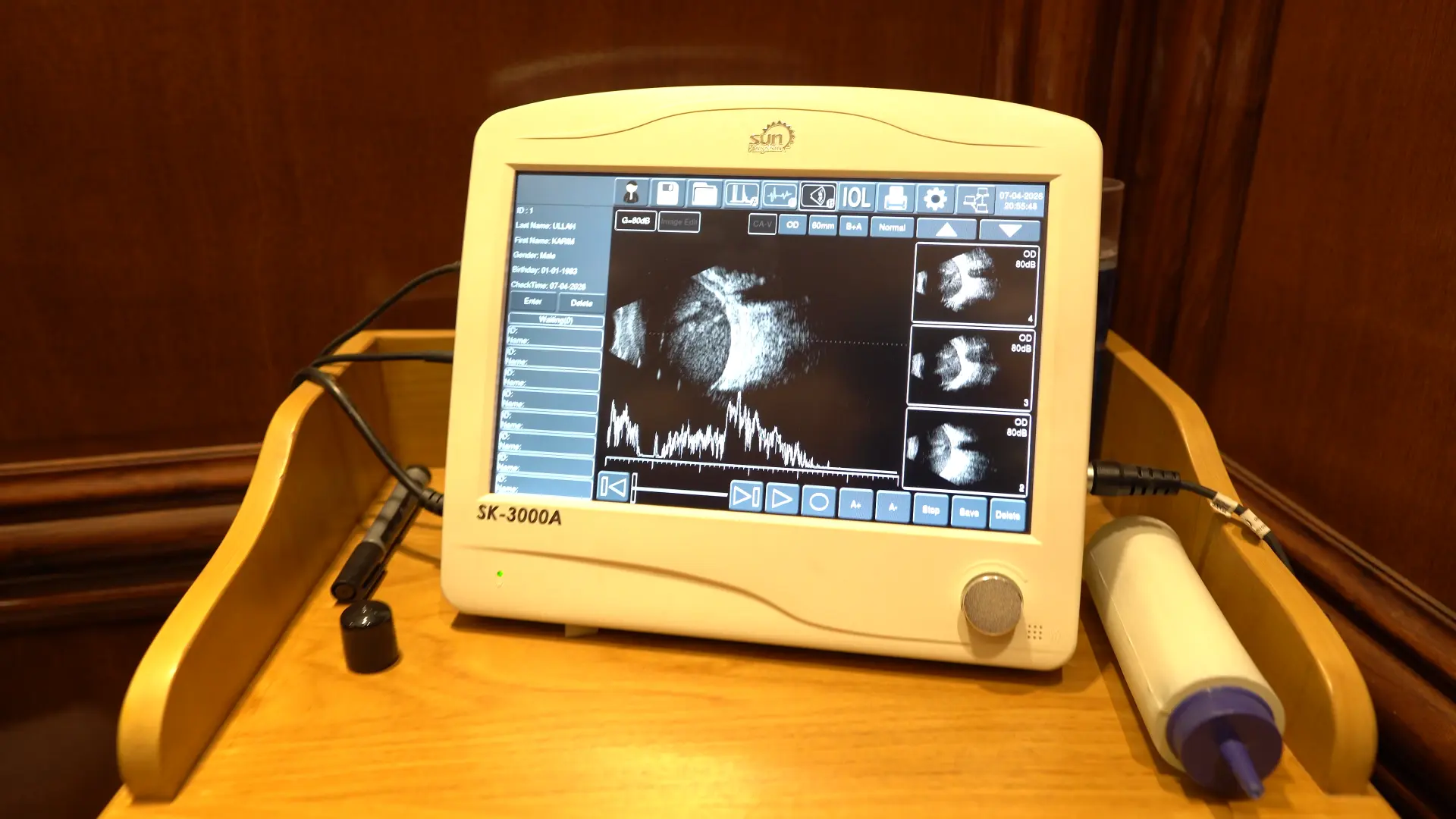
Ophthalmic A-scan and B-scan ultrasound unit imaging the retina through dense cataract in LahoreUnited States

Ophthalmic Ultrasound — A-Scan & B-Scan

Ultrasound lets us measure and examine the eye when the view is blocked by dense cataract or vitreous haemorrhage. A-scan supplies biometric measurement while B-scan images the retina and vitreous behind the opacity.

  • A-scan axial length measurement for lens power in dense cataract
  • B-scan imaging of retina and vitreous through opaque media
  • Detects retinal detachment and vitreous haemorrhage
  • Assessment and measurement of intraocular masses

Diagnostics Before Every Procedure

No one at IEH is booked for surgery before the measurements are in. Every treatment plan starts with the same four steps.

  1. 01

    Consultation

    A consultant reviews your history, symptoms and current prescription in detail.

  2. 02

    Corneal Mapping

    Corneal topography maps shape and thickness to confirm laser suitability.

  3. 03

    Retinal Imaging

    OCT and dilated examination check the macula, optic nerve and peripheral retina.

  4. 04

    Surgical Planning

    IOL Master biometry and measurements set your exact treatment or lens power.